Mesothelioma / Asbestos-Related Conditions / Pleural Plaques
Pleural Plaques and Asbestos Exposure
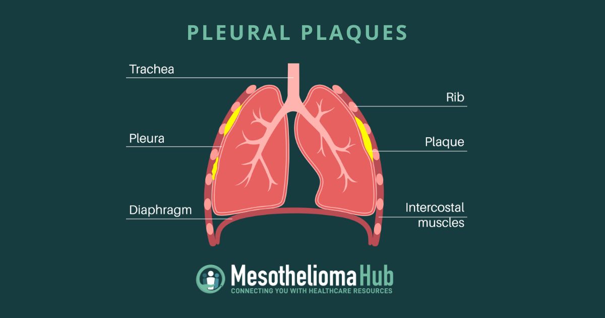
Pleural plaques are non-cancerous deposits of thickened tissue in the lining of the lungs. The plaques can occur from inflammation caused by asbestos exposure. Patients can experience pleural plaques 10 to 30 years after asbestos exposure.
Get Free Mesothelioma Guide
What Are Pleural Plaques?
Pleural plaques are non-cancerous collagen buildups that typically develop in the lining of the lungs. These are also an indicator that a person was exposed to asbestos and is at a greater risk for developing pleural mesothelioma or a different asbestos-related condition.
These plaques comprise fat, cholesterol, calcium, and other substances found in the blood. Plaques can occur in places outside of the lungs, such as in the arteries in patients with cardiovascular diseases. In some cases, plaque buildup is a likely cause of blood clots.
Prolonged asbestos exposure can cause the condition to develop in the membrane that surrounds the lungs and the chest cavity. Typically, these appear on scans more than 20 years after exposure, but rare cases have involved latency periods of less than ten years. Plaques located in the pleural area are not common in the general public, but there is a strong connection between pleural plaques and asbestos exposure.
Pleural Plaque Causes
The origin of pleural plaques occurs after someone is exposed to asbestos, typically associated with the rare and deadly disease called mesothelioma. Mesothelioma occurs after inhalation or ingestion of asbestos fibers that accumulate over time in the lining of the lungs. The buildup of fibers causes irritation and inflammation. The body’s response to this is to send fluid near the affected area. This fluid buildup causes pain and discomfort, and the healthy cells to mutate into cancerous ones.
However, this does not necessarily indicate that mesothelioma will occur, but a person with this as a result of asbestos exposure is at an increased risk for developing mesothelioma.
Similar to the development of mesothelioma, researchers believe the buildup of asbestos fibers causes an immune response. Instead of malignant cell mutation, the body creates pleural macrophage cells. Inflammation caused by the pleural macrophage cells causes thickening of the affected tissues, called fibrosis. The air sacs in the lungs normally appear long and thin but get thick and stiff with scar tissue over time. The scar tissues can calcify the pleural plaques on rare occasions.
Pleural Plaque Symptoms
Pleural plaques are asymptomatic, meaning they do not cause noticeable symptoms. A person can live with pleural plaques unknowingly until caught by chance. Over time, plaques may cause a slight decrease in lung function and capacity primarily due to pleural thickening.
Pleural thickening can cause symptoms of trouble breathing, chronic coughing, chest pain, or coughing up blood. Symptoms of pleural thickening can often indicate a more serious condition, such as cancer or asbestosis.
In some cases, a healthcare provider may recommend regular image tests if there is a known history of asbestos exposure among the patient. These imaging tests look for more serious conditions but often find the plaques.
Diagnosing Pleural Plaques
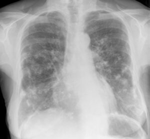
Since symptoms do not occur, a person is often diagnosed when undergoing radiography or CT imaging scans for other issues. The plaques typically occur in the parietal area of the pleura, which is the outer layer that lines the inner surfaces of the thoracic cavity. Plaques most often form in the lower portions of the chest.
X-Ray and CT Scans
X-ray imaging scans reveal grey-white areas of pleural thickening in the shape of a holly leaf. Most incidences of the plaques are found through x-ray scans. Calcified plaques can be difficult to identify through X-ray as they appear translucent or white in the lungs. A computerized tomography (CT) scan uses computer processing combined with X-ray images of different body angles to create cross-sectional images, or slices, of the bones, blood vessels, and soft tissues. Since calcified plaques can appear translucent, a CT scan can provide a more detailed image compared to an X-ray. Even if the plaques are not calcified, CT scans are the best way to diagnose this condition.
Treatment for Pleural Plaques
The condition does not typically require immediate treatment, is non-cancerous, and does not usually cause a loss in lung function. Doctors may recommend tests to observe the function of the lungs, the lung capacity, and how well oxygen reaches the bloodstream.
Patients with this condition should follow a healthier lifestyle to keep lung function strong and prevent further damage. While smoking does not cause asbestos-related issues, quitting can help the overall health of the lungs.
People with this condition should avoid further exposure to asbestos or use protective equipment such as respirators and protective clothing. Minimizing exposure to air pollution can help keep the lungs healthy and function properly. Avoid wildfires and areas with heavy air pollution.
Who Is at Risk for Developing Pleural Plaques?
Pleural plaques are not common among the general population. With less than 100,000 diagnoses each year, most cases result from workers exposed to asbestos on the job. Research shows that low levels of asbestos exposure can still cause this.
Occupations at risk or are related to prolonged asbestos exposure include:
- Asbestos miners and mining community members
- Automotive workers
- Construction workers
- First responders
- Shipyard workers
- Service members and veterans
If you were exposed to asbestos, inform your doctor about your exposure history and your risk for developing another asbestos-related condition or even worse.
Mesothelioma Support Team
Mesothelioma Hub is dedicated to helping you find information, support, and advice. Reach out any time!

